Guides & worksheets · Middle school: compare cell structures
Animal cell model project: compare animal and plant cell structures
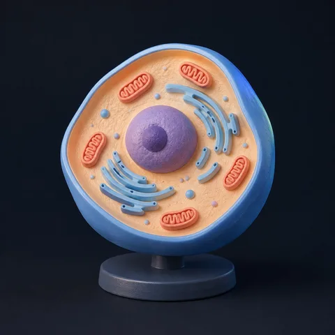
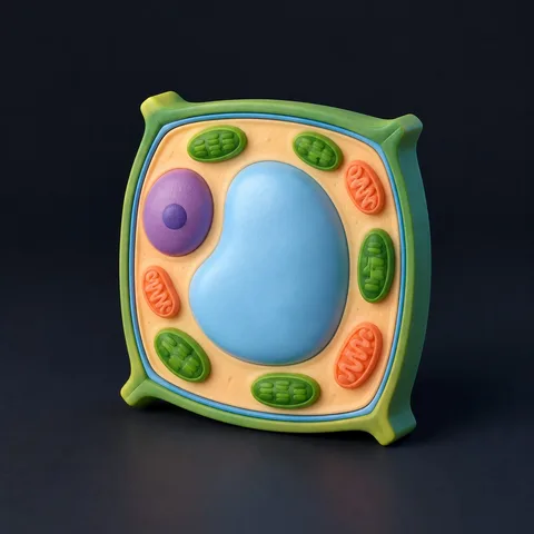
Start with the cell membrane, cytoplasm and nucleus shared by the typical cells shown here. Then compare the cell wall, chloroplasts and large central vacuole in the green plant cell example. Colors identify parts; they are not microscope colors or a universal picture of every cell.
By TangiSproutContent checked
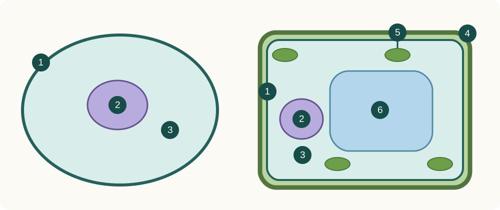
- Animal cell example
- Green plant cell example
- 1 Membrane
- 2 Nucleus
- 3 Cytoplasm
- 4 Cell wall
- 5 Chloroplast
- 6 Central vacuole
Find shared parts first
The diagram deliberately shows only a few structures. The cell membrane separates the inside from the surroundings and helps regulate exchange. The cytoplasm contains many cell components. The nucleus contains genetic material. Cell wall and cell membrane are different structures, not interchangeable labels.
Compare without overgeneralizing
The plant cell wall lies outside its membrane. This example represents a typical green plant cell: not every plant cell contains chloroplasts. Animal cells lack cell walls and chloroplasts, but it would be incorrect to conclude that they have no small vacuoles or vesicles.
Turn a model into an explanation
Choose one cell, label three structures and describe their relative positions. Explain one feature the model leaves out. A model can clarify an arrangement; it cannot replace microscopy or reproduce the scale, timing and full complexity of a living cell.
Observation and making activity
Learning resources
- Two pieces of card or paper for bases
- Paper shapes or modeling clay in three or more colors
- A pen and tape; scissors with adult help
- The diagram and your teacher’s required labels
- 1
Make an oval animal-cell base and a rounded rectangular green-plant-cell base. Keep a key so each color has the same meaning in both models.
- 2
Add membrane, cytoplasm and nucleus to both. On the plant model, add a separate wall outside the membrane, a large central vacuole and chloroplasts. Use paper shapes or small clay pieces.
- 3
Label each structure and compare the two models with the diagram. Explain one relationship the model shows and one it cannot show; add other structures only after checking your assignment and source.
Checks and explanations
- Membrane, cytoplasm and nucleus occur in the typical nucleated cells shown.
- The cell wall is outside the membrane; the green plant example includes chloroplasts and a large central vacuole.
- Teaching colors and proportions should not be treated as measured properties.
What the model can and cannot show
This is an original teaching schematic, not a micrograph or a medical atlas. AI reference images and generated models can contain errors; use the cited resources for factual checks.
Optional: create a digital model
You can finish the activity without generation. To explore another angle, create a digital model, then check its structures and omissions against the diagram.
Each live request generates one result and uses an allowance. Authorized sign-in is required.
Concept images are not finished models or verified structures. Choosing an image does not start generation.
Create model →